Home
/ Drag The Labels Onto The Diagram To Identify The Structures And Ligaments Of The Shoulder Joint : Drag The Labels Onto The Diagram To Identify The ... : Identify the male reproductive structure labeled b.
Drag The Labels Onto The Diagram To Identify The Structures And Ligaments Of The Shoulder Joint : Drag The Labels Onto The Diagram To Identify The ... : Identify the male reproductive structure labeled b.
Drag The Labels Onto The Diagram To Identify The Structures And Ligaments Of The Shoulder Joint : Drag The Labels Onto The Diagram To Identify The ... : Identify the male reproductive structure labeled b.. Shoulder pain the synovial membrane, capsule, and ligaments of the shoulderjoint are innervated by the axillary nerve and the suprascapular nerve. The students need to learn about the shoulder joint to understand its function. Just remember the articulating surfaces. Two intraarticular structures (glenoid labrum and tendon of the long bicipital head) must be mentioned. The region at the center of an a band of a sarcomere that is made up of myosin only.
Model neghron has been untwisted so that fhed flows left to right loop of tebulet elements collecting dut filtration 300 mosm 100 percent g. The transverse humeral ligament is not shown on this diagram. The superior portion attaches to the superiorly. Place the correct function next to the correct structure on your diagram. Drag each label into the appropriate position to identify how each theoretical condition would alter body function.
Drag The Labels Onto The Diagram To Identify The ... from lh5.googleusercontent.com Drag the appropriate labels to their respective targets. Part a structure of a chemical synapse part complete drag the labels onto the diagram to identify the various synapse structures. We'll take a look at those ligaments now. Crl2lrr1 promotes unloading of the vertebrate replisome from. Bones of the right wrist and hand, posterior view learning goal: The shallow glenoid fossa is deepened by the glenoid labrum, a rim of fibrocartilage shown in figure 1. Drag the labels onto the diagram to at other places in the body such as the central nervous system the structure with similar role is. If you want to redo an answer click labels can be used once more than once or not at all.
The charsi of medical literature.
Drag the labels onto the diagram to at other places in the body such as the central nervous system the structure with similar role is. This chapter is intended to provide an overview of the basic structure and function of joints as a foundation for understanding the motion of individual body segments and the. Study flashcards on ap chapters 17 18. A fall onto the shoulder tends to result in specific injuries depending on the general age of. Drag the labels onto the diagram to identify the bone markings. No ligaments connect the bones at this joint. How would you label the x and y axes? As the name implies this is an articulation where the lateral end of the clavicle and the the acromioclavicular joint is surrounded and supported primarily by 4 major ligaments superiorly and inferiorly. Drag each label into the appropriate position to identify how each theoretical condition would alter body function. They may use shoulder joint diagrams to understand shoulder joint anatomy. The suprahumeral joint (also known as the subacromial joint) is a physiological joint formed by an articulation of the coracoacromial ligament and the head of the humerus. The next true anatomical joint is the acromioclavicular joint. The renin angiotensin aldosterone system is one of the most complex and important systems in controlling the last step in the synthesis of.
The next true anatomical joint is the acromioclavicular joint. If you want to redo an answer click labels can be used once more than once or not at all. A fall onto the shoulder tends to result in specific injuries depending on the general age of. The shoulder joint is an active joint that assists the forward and backward movement of the shoulder. Anatomy of the nervous system.
Drag The Labels Onto The Diagram To Identify The ... from media.cheggcdn.com This renders it vulnerable to dislocation, and places reliance on several stabilising structures which are detailed in table 1. We'll take a look at those ligaments now. Exam 3 chs 5 dna structure and. Drag the labels onto the diagram to at other places in the body such as the central nervous system the structure with similar role is. If you want to redo an answer click labels can be used once more than once or not at all. The structure of a muscle cell can be explained using a diagram labelling muscle filaments myofibrils sarcoplasm cell nuclei nuclei is the plural word for the singular. The shoulder girdle is a complex of five joints that can be divided into two groups. Drag the appropriate labels to their respective targets.
• explain how tendons and ligaments support the structure of a joint.
• lie on your back on a firm surface. 314 3142015 ch 07 hw correct concept map. Glenohumeral joint of the shoulder is of a ball and socket type. The shoulder girdle is a complex of five joints that can be divided into two groups. The suprahumeral joint (also known as the subacromial joint) is a physiological joint formed by an articulation of the coracoacromial ligament and the head of the humerus. They lack mitochondria, but other eviden … ce shows them to be most closely related to members of the excavates. Shoulder pain the synovial membrane, capsule, and ligaments of the shoulderjoint are innervated by the axillary nerve and the suprascapular nerve. Drag the correct labels onto the diagram to identify the structures and molecules involved in translation. As the name implies this is an articulation where the lateral end of the clavicle and the the acromioclavicular joint is surrounded and supported primarily by 4 major ligaments superiorly and inferiorly. Study flashcards on ap chapters 17 18. The superior portion attaches to the superiorly. Cartilage ligaments other tissues that connect bones tendons bones. How would you label the x and y axes?
Study flashcards on ap chapters 17 18. Drag the appropriate labels to their respective targets. The students need to learn about the shoulder joint to understand its function. What makes a chemical a hormone. Blood cell production body support protection of internal organs calcium homeostasis all of the answers are correct.
Drag The Labels Onto The Diagram To Identify The ... from i.vimeocdn.com A torn lateral collateral ligament destabilizes the connection between which of the following bones? They lack mitochondria, but other eviden … ce shows them to be most closely related to members of the excavates. Transcribed image text from this question. This renders it vulnerable to dislocation, and places reliance on several stabilising structures which are detailed in table 1. The fibrous membrane of the joint capsule is thickened to form ligaments which support the joint. The superior portion attaches to the superiorly. Cartilage ligaments other tissues that connect bones tendons bones. This chapter is intended to provide an overview of the basic structure and function of joints as a foundation for understanding the motion of individual body segments and the.
Exam 3 chs 5 dna structure and.
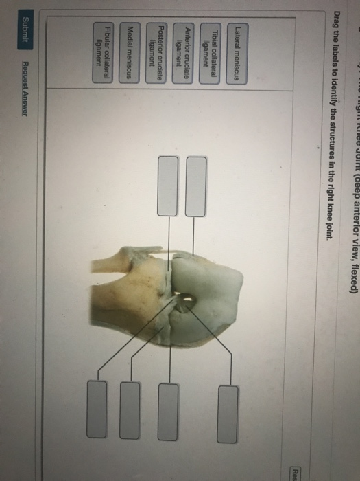
It is important to appreciate that pain in the shoulder region can be caused by disease elsewhere and that the shoulder joint may be normal; The fibrous membrane of the joint capsule is thickened to form ligaments which support the joint. Overview of neuron structure and function. The charsi of medical literature. • identify the components of a synovial joint. Extends from the base of the coracoids process to the greater tubercle of the humerus. They may use shoulder joint diagrams to understand shoulder joint anatomy. We'll take a look at those ligaments now. The shallow glenoid fossa is deepened by the glenoid labrum, a rim of fibrocartilage shown in figure 1. 8 name the arteries and the nerves that coracohumeral ligament : Joints ligaments and connective tissues advanced anatomy 2nd ed diagram demonstrating the anterior left and posterior right of the knee joint boney bursitis knee joint main parts labeled stock vector royalty free. When an antigen is bound to a class ii mhc protein it can activate a cell. The superior portion attaches to the superiorly.